Accurate coding for abdominal aortography and lower extremity angiography is one of the most technically demanding challenges in interventional radiology billing. These procedures vary in technique, access point, catheter positioning, and anatomical coverage, and each variable influences CPT code selection. A single documentation gap or misidentified catheter position can mean the difference between a clean claim and a denial.
In 2026, payer scrutiny on interventional radiology procedures has intensified. Medicare and commercial payers are tightening documentation requirements for RS&I codes, expanding prior authorization requirements for elective angiographic procedures, and increasing post-payment audit activity on catheter placement codes billed alongside imaging codes.
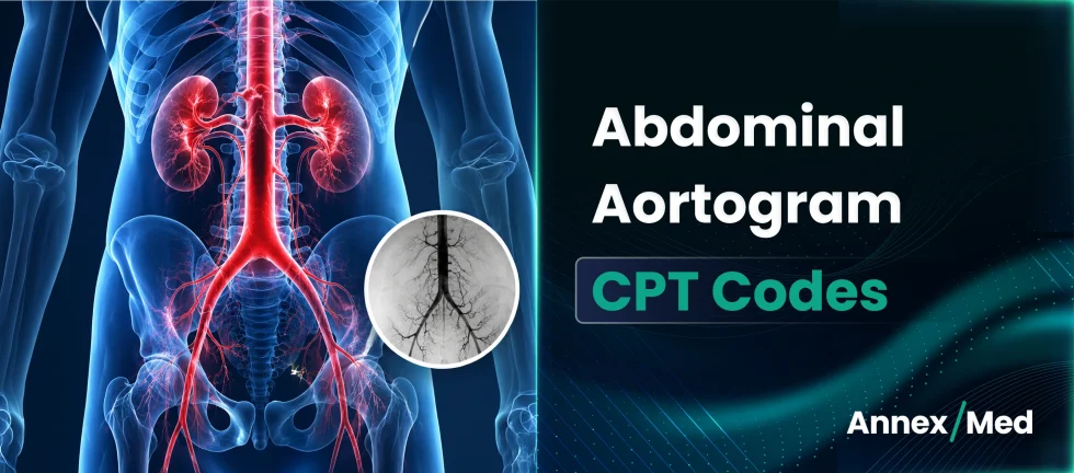
This guide covers the must-know CPT codes for abdominal aortography and angiography, anatomy context, RS&I codes, catheter placement codes, professional vs. technical component billing, modifiers, ICD-10 crosswalks, documentation standards, denial triggers, and the 2026 updates billing teams need to act on now.
Table of contents
- Aortic Anatomy and Its Impact on CPT Code Selection
- Interventional Radiology CPT Codes for Aortography
- CPT 75625 vs CPT 75630
- Bundling Rules and NCCI Edits for Aortography CPT Codes
- Modifiers in IR Billing
- ICD-10 Codes That Support Medical Necessity
- Documentation Requirements Per Code
- Turn Interventional Radiology Coding Precision Into Cleaner Claims
- FAQs
Aortic Anatomy and Its Impact on CPT Code Selection
Understanding vascular anatomy is essential for accurate interventional radiology coding. Catheter placement, imaging location, and runoff evaluation are all tied directly to the anatomy documented in the procedure note. The aorta is the largest artery in the human body, typically measuring around 2 to 3 centimeters in diameter. It is divided into four main sections. Each segment plays a distinct role in circulating blood and is often evaluated separately during imaging or interventional procedures.
Ascending Aorta – Emerges from the left ventricle. Relevant in thoracic aortic imaging but rarely the focus of peripheral vascular studies.
Arch of the Aorta – Curves left, ending at the level of T4–T5. Gives rise to the brachiocephalic, left common carotid, and left subclavian arteries.
Thoracic Aorta – Between the aortic arch and the diaphragm. Relevant when thoracic imaging accompanies abdominal studies.
Abdominal Aorta – Between the diaphragm and the common iliac arteries. The primary territory for abdominal aortography CPT codes. The renal arteries arise here whether the catheter is placed above or below the renal arteries is the critical coding distinction.
Coding implication: Catheter at or above the renal arteries → 75625 or 75630. Catheter repositioned below the renal arteries for extremity imaging → 75625 plus 75710 or 75716.
Catheter position relative to the renal arteries must be explicitly documented in every operative report since it is the single most important anatomical detail for aortography code selection.
Interventional Radiology CPT Codes for Aortography
There are specific CPT codes for imaging and catheter placement depending on how the procedure was performed. Coders must identify whether the catheter was placed in the aorta directly, repositioned, or remained in place during imaging. Similarly, they must determine which anatomical territories were imaged abdominal only or including lower extremities.
Radiology Supervision and Interpretation Codes:
CPT 75625 – Abdominal aortography by serialography, with radiological supervision and interpretation.
Used when a complete abdominal aortogram is performed with the catheter positioned at or above the renal arteries and no dedicated lower extremity runoff imaging is performed. Incidental visualization of the common iliac arteries does not convert this to 75630.
CPT 75630 – Abdominal aortography with bilateral iliofemoral runoff by serialography, with radiological supervision and interpretation.
Used when the catheter remains above the renal arteries and bilateral lower extremity arteries while imaging includes bilateral iliofemoral runoff without catheter repositioning.
CPT 75710 – Angiography, unilateral extremity, radiological supervision and interpretation
Used when imaging is performed on one lower extremity. Requires documentation confirming unilateral coverage and the extent of imaging at minimum to the knee.
CPT 75716 – Angiography, bilateral extremities, radiological supervision and interpretation
Used when both lower extremities are imaged. Documentation must confirm bilateral coverage to at least the knee level. If only one limb is imaged, use 75710.
Catheter Placement Codes:
CPT 36200 – Introduction of catheter into the aorta
The standard catheter placement code for femoral access with catheter advanced into the aorta. Reported in addition to the RS&I code not standalone.
CPT 36160 – Aortic, translumbar catheter placement.
Used when access is obtained directly through the lumbar approach rather than femoral. If a translumbar approach is used, report 36160 instead of 36200. They are not interchangeable based on access route.
RS&I codes and catheter placement codes are reported together. The RS&I code describes what was imaged and the catheter placement code describes how access was obtained. Both must be supported by the operative report.
Stop IR Denials Before They Reach Adjudication
AnnexMed helps IR practices strengthen coding accuracy, reduce denials, and improve reimbursement across complex angiography and aortography claims.
Request a Free Billing AssessmentCPT 75625 vs CPT 75630
The distinction between 75625 and 75630 is the most frequently searched and most frequently miscoded comparison in aortography billing. Both codes require catheter placement above the renal arteries where the difference is the extent of imaging performed.
Use CPT 75625 When:
- Catheter is placed at or above the renal arteries
- Complete abdominal aortogram is performed
- No imaging of the iliofemoral or extremity vessels
- Imaging of common iliac arteries is only incidental not a runoff study
Use CPT 75630 When:
- Catheter remains above the renal arteries
- Imaging includes bilateral iliofemoral arteries as a purposeful runoff study
- No catheter repositioning for leg imaging
- Both iliofemoral territories are covered in the same imaging sequence
When Neither Is Sufficient, Add Extremity Codes
If the catheter is repositioned to a lower position such as at the aortoiliac bifurcation to image the lower extremities separately, report:
- 75625 for the abdominal aortogram, plus
- 75716 for bilateral lower extremity imaging, or
- 75710 for unilateral lower extremity imaging
75630 is appropriate only when bilateral iliofemoral runoff is performed without catheter repositioning any repositioning for leg imaging requires 75625 plus a separate extremity angiography code.
Bundling Rules and NCCI Edits for Aortography CPT Codes
Bundling errors are the most common source of preventable denials in IR billing. Understanding which codes are included in others and which can be separately billed is essential for clean claim submission.
- Abdominal X-rays are bundled into RS&I codes: When 75625 or 75630 is reported, related abdominal X-rays (CPT 74018–74022) are considered bundled components and should not be separately billed. Reporting them alongside the RS&I code produces an automatic NCCI bundling denial.
- Bilateral requirement for 75716: To report 75716, documentation must confirm imaging of both lower extremities to at least the knee level. If only one limb was imaged or if bilateral imaging was performed but not documented, report 75710 instead. Reporting 75716 without bilateral documentation is an overcoding error that triggers audit review.
- 75630 and catheter repositioning: 75630 includes bilateral iliofemoral runoff without catheter repositioning. If the operative note documents catheter repositioning during the procedure, 75630 is not the correct code, report 75625 plus 75710 or 75716 based on the extent of extremity imaging.
- Catheter placement and RS&I codes: 36200 and 36160 are reported in addition to the RS&I codes. They describe catheter introduction, not imaging. Both components must be documented: the catheter placement method in the procedure note and the imaging interpretation in the radiology report.
Review NCCI edit tables for aortography code combinations at every quarterly update, bundling rules in IR change more frequently than most coders anticipate and missed updates produce systematic denials.
Clinical Scenarios for Accurate Coding
These scenarios help illustrate how code selection depends on catheter placement and imaging coverage.
Example 1 – Abdominal Aortography Only
- Access: Right femoral artery
- Catheter placed above renal arteries
- Imaging: Only abdominal aorta
- Codes: 36200, 75625
Note – If a translumbar approach was used, report 36160 instead of 36200
Example 2 – Aortography with Bilateral Iliofemoral Runoff
- Access: Right femoral artery
- Catheter placed above renal arteries
- Imaging: Abdominal aorta + bilateral iliofemoral vessels
- No catheter repositioning
- Codes: 36200, 75630
Example 3 – Aortogram Followed by Repositioning for Leg Imaging
- Access: Right femoral artery
- Initial catheter placement above renal arteries
- First, complete abdominal aortogram performed
- Then catheter moved to aortoiliac bifurcation for leg angiography
- Imaging includes bilateral lower extremities
- Codes: 36200, 75625, 75716
Note – If imaging was limited to one leg, replace 75716 with 75710
The operative report must document catheter position at each stage of the procedure such as initial placement, any repositioning, and the anatomical territory imaged at each position. Without this specificity, correct code selection cannot be supported at audit.
Modifiers in IR Billing
Precision in modifier use is essential for compliance and revenue protection.
| Modifier | Purpose | When to Apply | Denial Risk If Wrong |
| 26 | Professional component | Radiologist bills interpretation only, facility owns equipment | Overbilling with duplicate global claim |
| TC | Technical Component | Facility bills equipment and staff only | Underbilling or duplicate billing with global claim |
| 59 | Distinct procedural service | Two separately billable procedures on same date under NCCI rules | Overbundling procedures billed as one |
| 51 | Multiple procedures | Secondary procedure when billing multiple codes same session | Payer reduces secondary without modifier |
| RT/LT | Laterality | Unilateral extremity procedures | Missing laterality results in denial on Medicare claims |
Modifier 26 and TC split billing is the most common modifier error in interventional radiology confirm the practice setting and billing arrangement before every RS&I claim submission.
ICD-10 Codes That Support Medical Necessity
Proper ICD -10 selection validates medical necessity and strengthens claim defensibility during payer audits.
| CPT Code | Common ICD -10 | Description |
| 75625 | 171.4 | Abdominal aortic aneurysm without rupture |
| 75625 | I77.1 | Stricture of artery |
| 75630 | I70.0 | Atherosclerosis of aorta |
| 75630 | I74.09 | Other arterial embolism and thrombosis of abdominal aorta |
| 75710 | I70.213 | Atherosclerosis of native arteries of right leg with intermittent claudication |
| 75710 | I70.223 | Atherosclerosis of native arteries of left leg with intermittent claudication |
| 75716 | I70.203 | Atherosclerosis of native arteries of extremities, bilateral legs |
| 36200 | Reported with the imaging code diagnosis | Same diagnosis as the RS&I code on the claim |
ICD-10 must align with the clinical indication documented in the procedure report. A diagnosis-procedure mismatch is an independent denial trigger separate from any coding error.
Documentation Requirements Per Code
Operative and procedure reports must go beyond generic language to meet sufficiency standards. Detailed reporting ensures compliance, prevents denials, and supports medical necessity across all interventional radiology claims.
| CPT Code | What the Operative/Procedure Report Must Include |
| 75625 | Catheter position (at or above renal arteries), imaging coverage (abdominal aorta), no extremity imaging performed |
| 75630 | Catheter position (above renal arteries), bilateral iliofemoral runoff confirmed, no catheter repositioning |
| 75710 | Unilateral extremity specified, imaging extent documented (at minimum to knee), laterality confirmed |
| 75716 | Bilateral extremities confirmed, imaging extent to knee or below on both sides documented |
| 36200 | Femoral access documented, catheter advanced into aorta confirmed, approach type (femoral) stated |
| 36160 | Translumbar access documented, catheter placement in aorta confirmed, approach type (translumbar) stated |
Generic procedure language “aortogram performed” or “angiography completed” does not meet 2026 documentation sufficiency standards. Templates must prompt for catheter position, imaging territory, repositioning if any, and laterality on every IR procedure.
Turn Interventional Radiology Coding Precision Into Cleaner Claims
Interventional radiology comes with a unique set of coding and billing challenges. From detailed procedures like abdominal aortograms and runoff studies to complex catheter placements, accuracy matters, for getting reimbursed on time.
At AnnexMed, we bring that precision to the table. Our certified coders are trained to handle these complexities with care, making sure the right codes are used every time. We understand how one missed detail can lead to delays or denials. We help interventional radiology practices strengthen coding accuracy, reduce denials, and improve reimbursement through specialized IR billing expertise, certified vascular coders, NCCI edit compliance workflows, professional versus technical component billing support, and operative documentation review built specifically for complex angiography procedures.
If you’re looking for support that’s both reliable and experienced in IR billing, AnnexMed is ready to step in. We help simplify the process, reduce denials, and keep your cash flow steady.
Complex IR Procedures Need Specialized Billing Oversight
AnnexMed combines interventional radiology coding expertise, documentation review, and denial prevention workflows to support accurate reimbursement.
Talk to our Coding ExpertFAQs
- What is the difference between CPT 75625 and 75630?
CPT 75625 reports abdominal aortography without lower extremity runoff imaging, while CPT 75630 includes bilateral iliofemoral runoff imaging performed without catheter repositioning.
- Can CPT 75625 and 75716 be billed together?
Yes. CPT 75625 and CPT 75716 may both be reported when a complete abdominal aortogram is performed and the catheter is repositioned for separate bilateral lower extremity angiography.
- When should CPT 75710 be reported?
CPT 75710 is used when angiographic imaging is performed on only one extremity rather than both lower extremities.
- Is catheter repositioning required for CPT 75630?
No. CPT 75630 applies when bilateral runoff imaging is performed without repositioning the catheter from its original placement above the renal arteries.
- Are abdominal X-rays separately billable with 75625 or 75630?
No. Routine abdominal imaging performed during angiography is generally bundled into the RS&I angiography CPT codes and should not be billed separately.
- Why do angiography claims get denied?
Common denial reasons include incomplete runoff documentation, incorrect unilateral or bilateral coding, missing catheter positioning details, and billing bundled imaging services separately.